Evisceration is an ophthalmic surgery in which the internal contents of the eye is removed leaving the scleral shell and extraocular muscles intact.
Usually, it is followed by placement of an orbital implant to replace the lost ocular volume.
Some facts about Evisceration:
- Usually, Evisceration is performed to reduce pain or improve cosmesis in a blind eye.
- An ocular prosthetic can be fitted after the surgery over the eviscerated eye in order to improve cosmesis.
- All intraocular contents are removed while preserving the remaining scleral shell, extraocular muscle attachments and surrounding orbital adnexa in Evisceration.
- Placement of an implant into the evisceration cavity is oten included in the surgery to maintain appropriate orbital volume.
- Endophthalmitis, penetrating ocular trauma, blind, painful eye are some of the indication of Evisceration.
- Due to a lack of sufficient scleral shell volume to adequately encase the implant, Evisceration may be more difficult in cases of phthisis bulbi or microphthalmos.
- Retrobulbar hemorrhage, Orbital edema, Dissemination of unexpected intraocular neoplasm, Implant exposure and Implant extrusion are some of the complications of Evisceration.
Procedure for Evisceration:
- A pre-operative evaluation should be performed to ensure there is no intraocular malignancy in the operative eye when planning for an evisceration.
- B-scan or CT scan should be used to rule out malignancy if there is no view to the posterior pole.
- Enucleation is the preferred surgery if intraocular malignancy can not be ruled out.
- Correct laterality of the evisceration must be confirmed before starting the procedure.
- Evisceration is mostly performed under general anesthesia. However, intravenous, monitored sedation with or without regional blocks can be used in some cases.
- Anesthetic with epinephrine is often given to reduce intraoperative bleeding and postoperative pain.
- Local anesthetic may be injected subconjunctivally to distinguish available conjunctiva and Tenon's capsule and aid in hemostasis.
- Several drops of 10 percent topical phenylephrine can also be applied to the ocular surface before the procedure to provide hemostasis.
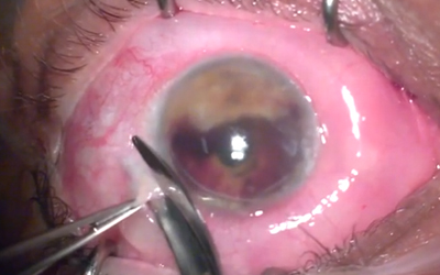
- A 360 degree conjunctival peritomy is made at the limbus to undermine the conjunctiva and Tenon's capsule in a careful anterior dissection before utilizing Wescott scissors.
- Scissors may be introduced to excise the cornea in a circumferential manner by making a full thickness incision at the limbus.
- All intraocular contents, including uveal tract, vitreous humor, crystalline lens and retina are then removed.
- An evisceration spoon, spatula, suction, or other instruments can be used for removing the intraocular contents.
- These contents are sent for examination and histopathologic identification.
- A trap should be placed to retain the contents for histopathology if suction is used.
- Cautery and direct pressure is then used to achieve the hemostasis of the nerve and vortex veins.
- All remaining uveal material and microorganisms will be removed from the scleral shell by using absolute or 70 percent alcohol.
- Care must be taken to keep the alcohol within the sclera and not contact the conjunctiva as additional irritation and edema may be created by this practice.
- A posterior sclerotomy or similar scleral relaxing incisions will be made to keep ready the evisceration cavity and allow for a larger implant to be placed in some cases.
- The best implant size will be evaluated and chosen by the surgeon to restore orbital volume while ensuring appropriate position.
- The scleral shell is further bathe in antibiotic solution prior to implant placement by some surgeon.
- The material or implant is surgeon dependent and includes spherical implant choices of acrylic, silicone, PMMA, porus polyethylene and hydroxyapatite.
- The implant may be either placed directly into the scleral shell or may be first wrapped in donor sclera, mesh or other materials
- Opening the posterior sclera, releasing the optic nerve, and placing the orbital implant behind the scleral shell, and closing a double layer of sclera over the anterior implant are some of the other techniques.
- Placement of a large orbital implant is allowed which decreases superior sulcus hollowing and anophthalmic ptosis, which also results in a better cosmetic result.
- The anterior sclera, Tenon's capsule and conjunctiva is then closed carefully in a layered approach before placement of a conformer.
- A temporary tarsorrhaphy can also be performed to help the conformer remain in place.
- Perioperative and/or postoperative antibiotics are often administered after the procedure. These are especially important in cases of evisceration in the setting of endophthalmitis.
- The duration of antibiotic therapy ranges from 10 days to several weeks, depending on the nature of the infection.
- Surgical instruments and gloves are generally exchanged by the surgeon prior to implant placement and closure to lessen the risk of contamination and prolonged infection.
- An additional retrobulbar administration of anesthetic or ethanol can be placed prior to patching to manage post-operative pain.
- A pressure patch may be applied and kept in place for approximately five days following surgery.
- Prosthesis fitting can be done 6 to 8 weeks following surgery.
- The implant can be placed in a secondary staged procedure days to weeks after the initial evisceration. This is particularly in case of acute infection, as that the rates of implant extrusion may be higher with primary implantation at the time of evisceration
- The use of a dressing, pressure patch, and/or ice cold compresses are common post-operative care instructions to help with post-operative edema and comfort
Special Considerations:
- Scleral buckle or glaucoma drainage device should be removed, if present in the eviscerated eye.
- The limbus can be incised to irrigated the silicone oil from the eye prior to peritomy if it is present within the eye.
